K-SCAN (Iopamidol Injection)
K-Scan (Iopamidol Injection) is a trusted non-ionic contrast medium that has become a staple in modern radiology for its balance of diagnostic efficacy and patient safety. Engineered to be highly water-soluble, K-Scan provides excellent opacity for a broad spectrum of X-ray and CT procedures. Its formulation is designed to minimize physiological disruption, ensuring high-quality images while maintaining a safe profile for diverse patient populations, from neonates to the elderly.
Overview
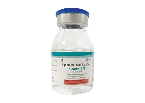
K-Scan is available in two primary concentrations—300 mg/ml and 370 mg/ml—to meet the specific needs of various imaging protocols. The 370 mg/ml concentration is particularly effective for high-flow vascular studies and for patients with mild to moderate renal insufficiency, as it induces less osmotic diuresis than ionic alternatives. This reduced osmotic load is a critical trust signal for clinicians managing sensitive cases, including neonatal and geriatric patients, ensuring diagnostically useful nephrography even in those with severe renal impairment.
Key Features
- Non-Ionic Formulation: Significantly reduces the risk of adverse reactions compared to older ionic contrast agents.
- Low Viscosity & Osmolality: Optimized viscosity (9.4 mPa.s at 37°C for the 370 concentration) facilitates smooth injection and improved patient tolerance.
- Excellent Opacification: Provides high-density imaging for precise visualization of the vascular system and urinary tract.
- Renal Tolerance: Specifically noted for its suitability in patients with renal insufficiency due to its balanced osmotic profile.
Clinical Applications
- Angiography & Angiocardiography: Used for cerebral, coronary, thoracic, and abdominal aortography to visualize steno-occlusive diseases.
- Cath Lab procedures- for Angiography and Angioplasty it is a drug of choice.
- Urography: Standardized for intravenous urography in adults and children to assess renal and urinary tract function.
- CT Enhancement: Improves the visualization of internal organs and structures during computerized axial tomography (C.T.) scans.
Inquiry Form